Aortic Valve Disease
Aortic valve disease is a cardiovascular condition caused by alterations in the effectiveness of the valve between the heart’s left ventricle and a large artery called the aorta.
About Aortic Valve Disease
Your left ventricle pumps oxygenated blood into your body through a large artery known as the aorta. The aortic valve is the structure that regulates the flow of blood from the left ventricle to the aorta. When the left ventricle contracts to pump blood, the aortic valve opens, allowing blood to pass through. As the left ventricle relaxes, the aortic valve closes to prevent blood flow back into the ventricle (regurgitation). Aortic valve disease is a form of valvular heart disease caused by irregularities which disrupt the proper opening and closing of this aortic valve and subsequently the delivery of oxygenated blood to your heart, your head, and the rest of your body. While your symptoms may vary, some untreated aortic valve disease can result in serious problems, making it important to address and monitor your condition over time. Additionally, aortic valve disease can change over time so it is important to have routine check ups as suggested by your doctor.
Types of Aortic Valve Disease
Aortic valve disease is classified by which of the aortic valve’s functions is affected; it is possible to experience a combination of both forms of the condition.
Types of aortic valve disease include:
- Aortic stenosis: Constriction or narrowing of the aortic valve which prevents adequate passage of blood from the left ventricle to the aorta.
- Aortic regurgitation: Improper closure of the aortic valve which allows blood to flow backwards from the aorta into the left ventricle.
Symptoms of Aortic Valve Disease
Symptoms of aortic valve disease vary, and some patients do not experience any symptoms.
Symptoms of aortic valve disease may include:
- Chest pain
- Dizziness
- Heart murmur
- Lack of energy
- Shortness of breath
- Sleep difficulty
- Swelling feet or ankles
Risk Factors for Aortic Valve Disease
Certain patients are at higher risk for aortic valve disease.
Risk factors for aortic valve disease may include:
- Age: Increased age is associated with increased wear of the aortic valve tissue, which may result in aortic valve disease.
- Health history: Certain birth defects can cause aortic stenosis, even in young people, while infection, injury, and high blood pressure are linked to aortic regurgitation.
- Congenital abnormalities: A bicuspid aortic valve or other structural abnormalities present at birth can contribute to aortic valve disease, though many patients do not learn about these defects until later in life.
- Genetics: Certain genetic conditions, such as Marfan Syndrome, Loeys-Dietz Syndrome, and other connective tissue disorders, can increase the risk of developing aortic valve disease.
Diagnosing Aortic Valve Disease
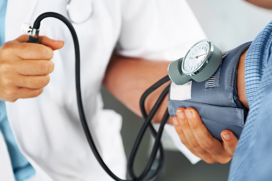
After performing a physical examination and assessing your medical history, your doctor may suggest additional testing if they suspect you have aortic valve disease.
Tests performed when diagnosing aortic valve disease may include:
- Cardiac Catheterization: During cardiac catheterization, a small catheter (thin tube) is inserted into a larger blood vessel, typically in the groin, and guided to the heart where blood pressure and oxygen measurements can be taken in the aorta and pulmonary artery as well as the four chambers of the heart. A dye can also be injected through the tube to make the heart’s structure more visible on an X-ray
- Chest X-Ray: A chest X-ray produces an image of the tissue and bones in the heart and lungs and helps your provider assess the shape, size, and structure of the heart and lungs as well as the aeration of or any congestion in the lungs.
- Echocardiogram: The most common diagnostic test for valvular heart diseases, an echocardiogram uses ultrasound technology to create a moving image of the heart and its valves, allowing your provider to assess the structure and function of the heart. An echocardiogram also helps provide information about blood flow and how well the heart is pumping blood.
- Electrocardiogram (ECG or EKG): An electrocardiogram uses electrodes that are placed on the body to record the electrical activity taking place in the heart. An ECG/EKG test helps detect abnormal rhythms, such as cardiac arrhythmias, stress on the heart, and damage to the heart muscles.
- Stress Test: During a stress test, a doctor monitors your heart function and blood pressure while you perform exercise.
Treating Aortic Valve Disease
Treatment for aortic valve disease may vary depending on your form of the condition, the severity of your symptoms, and your overall health. Your care team will work with you to determine the best course of treatment.
Treatment options for aortic valve disease may include:
- Balloon Valvuloplasty: Using the cardiac catheterization method, a small tube can be inserted through a blood vessel in the groin and guided to the heart. A balloon can be pushed through the tube and inflated in the aortic valve to stretch the narrow portion. A stent may be added after the balloon is removed to ensure the aorta stays open.
- Valve Replacement: The aortic valve can be surgically replaced with a biological valve made from animal tissue or a mechanical valve. The pulmonary valve and a portion of the pulmonary artery from the patient (Ross procedure) or an aortic valve from a donor can also be used to replace the valve and part of the aorta. This procedure can be performed using either open-heart surgery or minimally invasive surgery known as transcatheter aortic valve replacement (TAVR), which involves accessing the valve via an artery in the groin under monitored anesthesia.
- Valve Repair: This includes procedures like a valvotomy, where the valve is surgically altered to improve function by cutting or removing parts of the valve tissue that are causing obstruction.
Treatment options for DORV may include:
- Arterial Switch: A type of surgery in which the position of the arteries are reversed so that the pulmonary artery is connected to the right ventricle and the aorta is connected to the left ventricle. If the child also has a ventricular septal defect, this is typically closed by placing a patch over the hole during the same operation.
- Intraventricular Repair: A type of surgery in which a tunnel (baffle) is created through the ventricular septal defect to connect the aorta to the left ventricle.
Care Team Approach
The Institute for Cardiovascular Health care team treats all types of heart disease and vascular disorders. Patients are cared for by a dedicated multidisciplinary care team, meaning you will benefit from the expertise of multiple specialists across a variety of disciplines. Our board-certified and fellowship-trained heart surgeons have extensive experience treating heart disease and vascular disorders and work alongside a team of cardiac experts, including cardiologists, advanced practice providers, and more, providing unparalleled care for patients every step of the way.
We collaborate with our colleagues at the Dell Medical School and The University of Texas at Austin to utilize the latest research, diagnostic, and treatment techniques, allowing us to identify new therapies to improve treatment outcomes. We are committed to communicating and coordinating your care with referring physicians and other partners in the community to ensure that we are providing you with comprehensive, whole-person care.
Learn More About Your Care Team
Institute for Cardiovascular Health
Ascension Seton Medical Center - Main
1201 W. 38th Street, Austin, TX 78705
1-512-324-3028
Get Directions